Ureter Course In Female Pelvis
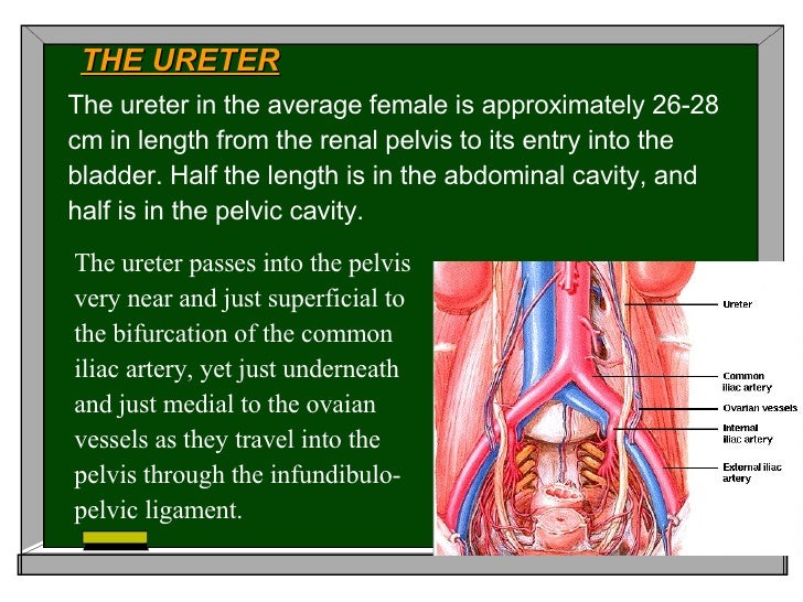
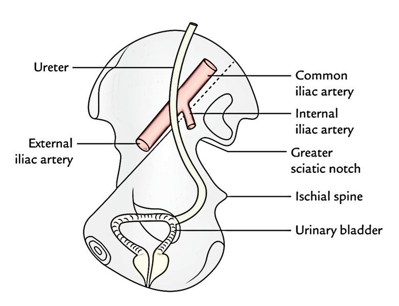
Ureter Course In Female Pelvis - The urethra is a part of the renal system, which also includes the kidneys, ureters, and the bladder. From there, these muscular tubes travel along the pelvis' lateral wall and connect to the urinary bladder. From the renal pelvis to the pelvic brim. It is a funnel shape upper expansion of the ureter. It begins at the neck of the bladder, traverses the pelvic and urogenital diaphragms, and ends at the external urethral orifice. The ureters are a pair of muscular tubes which convey the urine from kidneys (renal pelvis) to the urinary bladder. In the pelvis, the ureter first runs downward, backward, and laterally along the anterior margin of the greater sciatic notch. The female urethra, about 4 cm in length, is fused with the anterior wall of the vagina. The ureters travel inferiorly from the renal pelvis apices at the kidney hila, pass anterior to the psoas, and course over the pelvic brim at the common iliac artery bifurcation. In the majority of the patients, the course of the ureter is easily demarcated from the level of the pelvic brim. The urethra is a fibromuscular tube that conducts urine from the bladder (and semen from the ductus deferens) to the exterior. It may lie completely outside the kidney or buried inside the substance of the renal hilum. In this zone, the ureter travels medial and inferior to the gonadal vessels and enters the pelvis by crossing over the common iliac vessels at the bifurcation. Kidneys and ureters in cadavers: The female urethra starts at the base of the bladder and continues down through the pelvic floor. Additionally, a child with dv may experience storage symptoms such as frequency and. They begin at the ureteropelvic junction, where the renal pelvis continues on as the ureter. It then runs medialward and forward on the lateral aspect of the cervix uteri and upper part of the vagina to reach the fundus of the bladder. See section trigone of the urinary bladder for the anatomy of the ureteral orifice. In the female, the ureters pass under the ovarian and uterine vessels. In this zone, the ureter travels medial and inferior to the gonadal vessels and enters the pelvis by crossing over the common iliac vessels at the bifurcation. The urethra is a fibromuscular tube that conducts urine from the bladder (and semen from the ductus deferens) to the exterior. Retroperitoneal structure in the posterior abdominal wall (upper part) and lateral pelvic. Opposite to the ischial spine, it turns forwards and medially to get to the base of the urinary bladder, where it enters the bladder wall obliquely. Its upper half courses in the abdomen (abdominal part) while its lower half courses in the pelvis (pelvic part). In the pelvis, the ureter first runs downward, backward, and laterally along the anterior margin. The ureters are muscular tubes that run from the kidneys to the urinary bladder. Congenital anomalies of the pelvic ureter important for gynecologist: Dysfunctional voiding (dv) is a multifactorial functional problem that refers to dysfunction during voiding. (1) ectopic ureter that opens in the vestibule, urethra, vagina or cervix. Opposite to the ischial spine, it turns forwards and medially to. From the pelvic brim to the bladder. In the pelvis, they receive additional branches from the internal iliac, middle rectal, uterine, vaginal, and vesical arteries. In women, the ureter lies dorsally of the round ligament, uterine artery and above mentioned structures. The urethra is a fibromuscular tube that conducts urine from the bladder (and semen from the ductus deferens) to. Opposite to the ischial spine, it turns forwards and medially to get to the base of the urinary bladder, where it enters the bladder wall obliquely. Ureter is the canal through which urine is transported from the kidney to the bladder. In the female, the ureters pass under the ovarian and uterine vessels. Retroperitoneal structure in the posterior abdominal wall. Each one has a length of 30 centimeters (approximate), which advance from the bottom of each kidney, following through the lower abdomen and the pelvis first area. From the pelvic brim to the bladder. The upper ureter, zone 1, is the portion extending from the renal pelvis to iliac arteries. During their course in the abdomen, the ureters receive blood. Dv is clinically important because it increases the risk of urinary tract infections, mostly due to incomplete bladder emptying, and unfavorably affects renal function. In general the ureter is seen crossing the external iliac vessels from lateral to medial at the base of the infundibulopelvic ligaments. In the abdomen the branches arise medial to the ureter and in the pelvis,. It is a funnel shape upper expansion of the ureter. In this zone, the ureter travels medial and inferior to the gonadal vessels and enters the pelvis by crossing over the common iliac vessels at the bifurcation. Ureters are continuations of the renal pelvis, which is located posterior to the renal artery and renal vein (acronym 'avp'). Each one has. From the ischial spine, it turns forwards and medially to reach the superolateral angle of the base of urinary bladder, where it enters the bladder wall. Additionally, a child with dv may experience storage symptoms such as frequency and. From the renal pelvis to the pelvic brim. In this zone, the ureter travels medial and inferior to the gonadal vessels. In the pelvis, they receive additional branches from the internal iliac, middle rectal, uterine, vaginal, and vesical arteries. Additionally, a child with dv may experience storage symptoms such as frequency and. In the pelvis, the ureter first runs downward, backward, and laterally along the anterior margin of the greater sciatic notch and reaches the level of ischial spine. In both. Retroperitoneal structure in the posterior abdominal wall (upper part) and lateral pelvic wall. Dv is clinically important because it increases the risk of urinary tract infections, mostly due to incomplete bladder emptying, and unfavorably affects renal function. In the pelvis, the ureter first runs downward, backward, and laterally along the anterior margin of the greater sciatic notch and reaches the level of ischial spine. The ureters travel inferiorly from the renal pelvis apices at the kidney hila, pass anterior to the psoas, and course over the pelvic brim at the common iliac artery bifurcation. From there, these muscular tubes travel along the pelvis' lateral wall and connect to the urinary bladder. It is a funnel shape upper expansion of the ureter. They begin at the ureteropelvic junction, where the renal pelvis continues on as the ureter. Kidneys and ureters in cadavers: From the pelvic brim to the bladder. It begins at the neck of the bladder, traverses the pelvic and urogenital diaphragms, and ends at the external urethral orifice. Gynecologic and urologic surgery is frequently performed using a vaginal or perineal approach. Ureter is the canal through which urine is transported from the kidney to the bladder. The ureters can be confused with the inferior mesenteric artery. The distinguishing feature is that the ureter passes posterior to the vessel. The female urethra, about 4 cm in length, is fused with the anterior wall of the vagina. Its upper half courses in the abdomen (abdominal part) while its lower half courses in the pelvis (pelvic part).
Cardinal Ligament Ureter
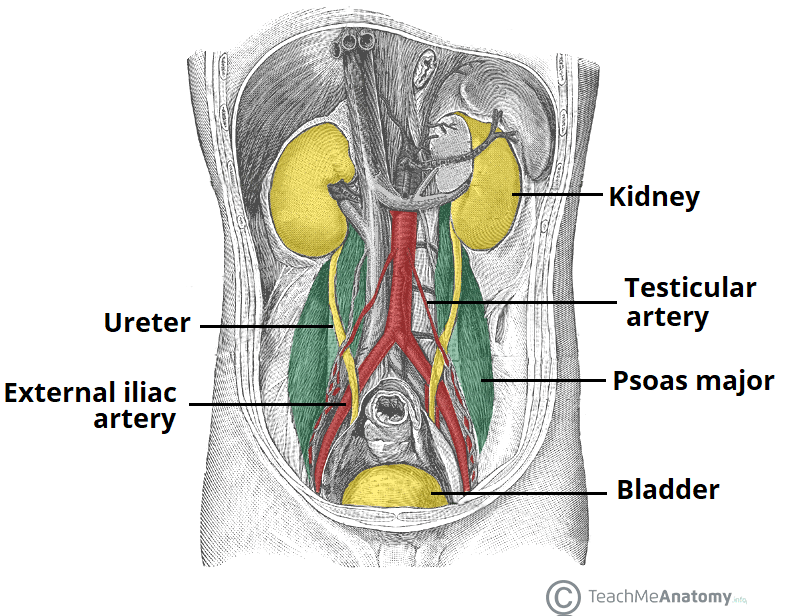
The Ureters Anatomical Course Neurovascular Supply TeachMeAnatomy

Female Pelvic Anatomy Ureter ANATOMY STRUCTURE
Anatomy2009
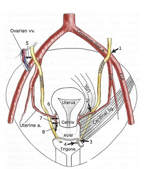
Course of pelvic ureters. Taken from [1]. Download Scientific Diagram

Anatomy of the Female Urinary Tract Obgyn Key
Pelvic ureter
Ureter Earth's Lab
مركز صحة المرأة والتعليم إصابة المسالك البولية الوقاية والإدارة

Course of ureter female Diagram Quizlet
In This Zone, The Ureter Travels Medial And Inferior To The Gonadal Vessels And Enters The Pelvis By Crossing Over The Common Iliac Vessels At The Bifurcation.
It May Lie Completely Outside The Kidney Or Buried Inside The Substance Of The Renal Hilum.
In The Pelvis, The Ureter First Runs Downward, Backward, And Laterally Along The Anterior Margin Of The Greater Sciatic Notch.
The Ureter Begins Its Descent To The Bladder By Running Along The Medial Aspect Of The Psoas Muscle.
Related Post: